
Figure d'article scientifiqueGiant viruses of the polar regions: diversity, endemism, adaptation and ecological structuring
Thomas M. Pitot , Catherine Girard (2026)
Université Laval.
Thomas M Pitot, Catherine Girard, Giant viruses of the polar regions: diversity, endemism, adaptation and ecological structuring, FEMS Microbiology Ecology, 2026;, fiag061, https://doi.org/10.1093/femsec/fiag061
© The Author(s) 2026. Published by Oxford University Press on behalf of FEMS.
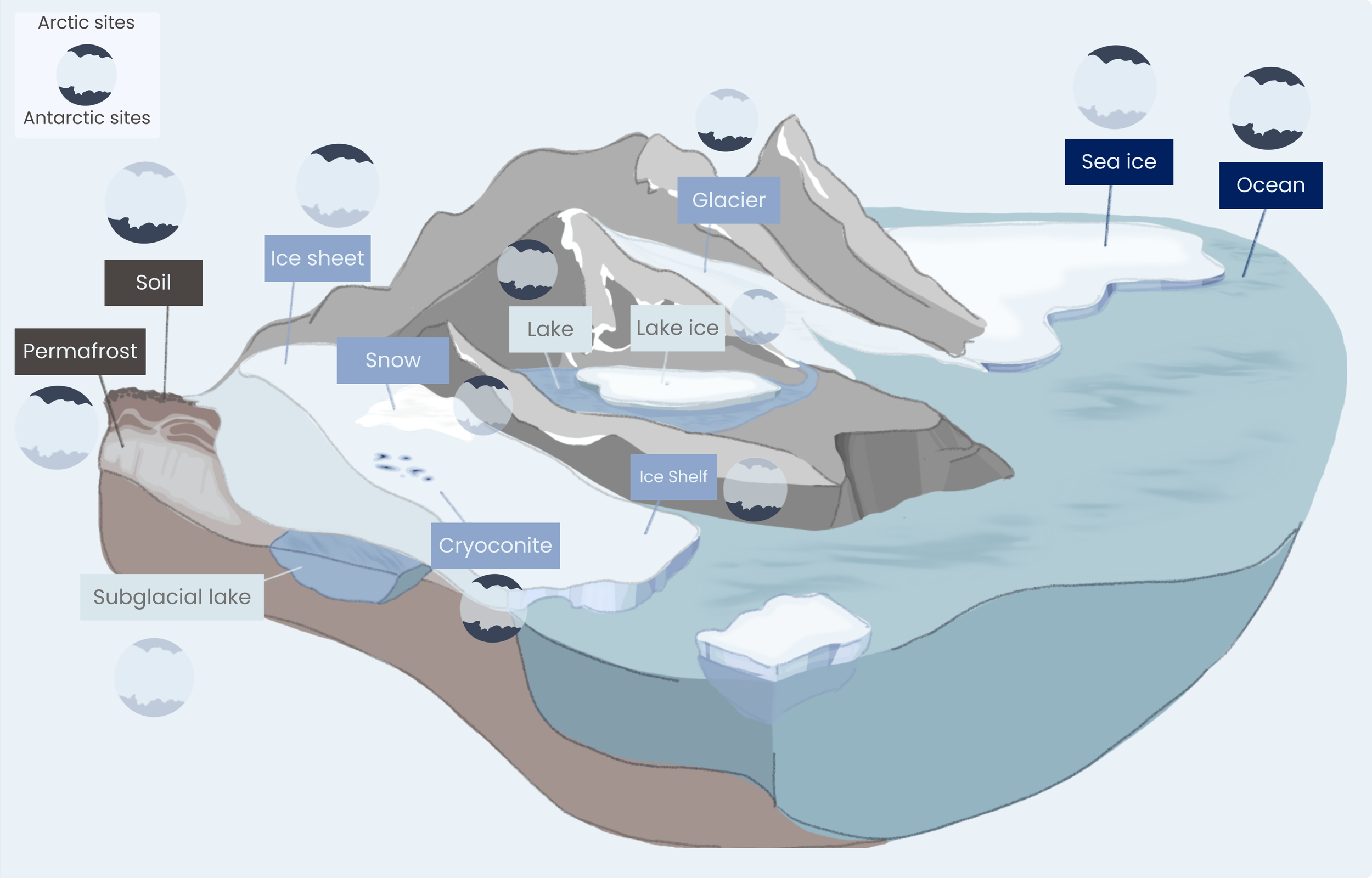
Figure 1: Schematic representation of polar cryospheric marine and terrestrial habitats and systems, with graphical markers indicating the polar region in which giant viruses have been reported to date (Arctic, Antarctic, or both). The schematics are intended solely for visualization purposes and do not aim to provide a high‑fidelity representation of real ecosystems.
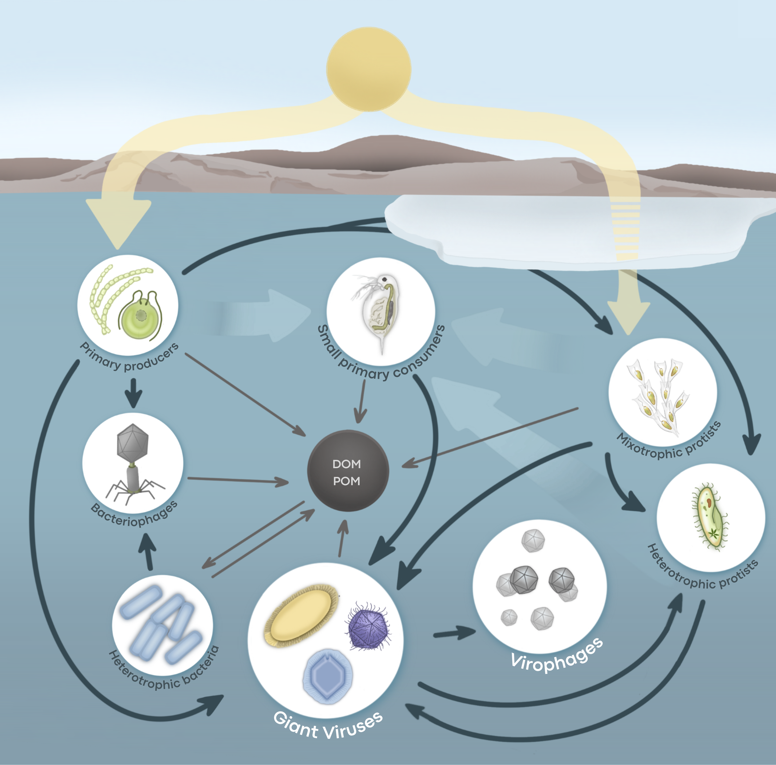
Figure 2: Microbial and viral trophic web structure in polar lakes. Multicellular organisms and higher trophic levels are rare or absent, resulting in a truncated food web dominated by unicellular organisms and viruses. Dark blue arrows show predation, parasitism and infections relationships. Yellow arrows show light penetration in water and through ice. Pale arrows represent higher trophic relationship, and brown arrows show the release of organic matter release. The schematics is intended for visualization purposes; organisms are not to scale, and do not represent any specific species.
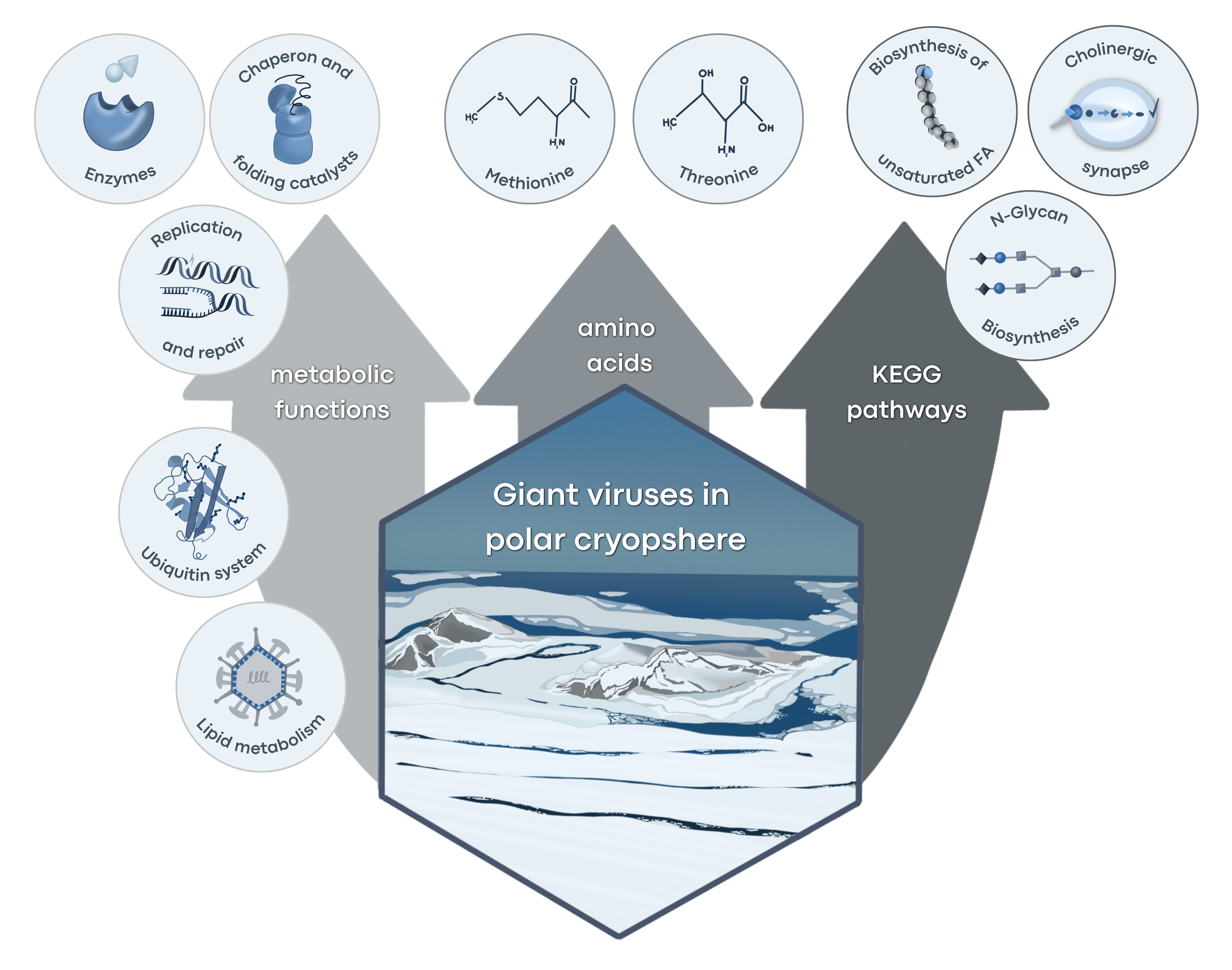
Figure 3: Schematic representation of polar‑speciϐic or polar‑enriched giant viral amino acid composition, metabolic functions and pathways in cryosphere giant viruses. The visualizations are primarily inspired by data and results from cold‑adaptation studies by Meng et al. (2023) and Buscaglia et al. (2024). These schematics are intended solely for visualization purposes.
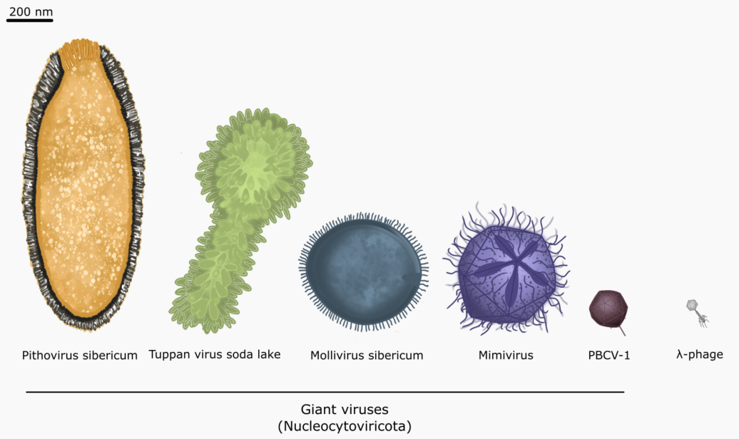
Figure 4 : Schematic representation of size and shape variation between selected Nucleocytoviricota virions and lambda phage for scale. From left to right; Pithovirus sibernicum (∼1.5 µm in length, 500 nm in diameter), Tupanvirus soda lake (∼1 µm in length, capside ∼450 nm in diameter, tail ~550 nm extension), Mollivirus sibernicum (~550 nm in diameter), Mimivirus bradfordmassiliense (AMPV) (~550 nm in diameter, ~750 nm with ϐibrils), Chlorovirus PBCV-1 (~190 nm in diameter), Lambda phage (capside ~50-65 nm in diameter, ~200 nm in length). These schematics are intended solely for visualization purposes.